Saturday, October 31, 2020

Lupine Publishers| Multidisciplinary foot Clinic Reduces Diabetic Lower Limb Amputation but Cannot Prevent it Completely: 13 Year Experience at a Vascular Tertiary Centre

 Lupine Publishers| Multidisciplinary foot Clinic Reduces Diabetic Lower Limb Amputation but Cannot Prevent it Completely: 13 Year Experience at a Vascular Tertiary Centre

Lupine Publishers| Diabetes and Obesity

Abstract

Objectives: Assess the effect on lower limb major amputation rate following the introduction of the multi-disciplinary foot clinic in a defined relatively static population in the North West of England.

Methods: Data surrounding all lower limb amputations in diabetic patients between 1997 and 2010 were retrospectively collected and analysed.

Results: Results demonstrated significant increase in minor amputations but numerical reduction in major amputations. There was significant change in ratio of minor to major amputation. When annual fluctuation was averaged out over 5-year period, the annual major amputation per 100,000 patients reduced from 6.6 to 5.1 with increase in minor amputations from 2.0 to 5.9.

Discussion: A reductions in major amputations rate with increase in minor amputations occurred over the 13-year period following implementation of the Multi-Disciplinary Foot Clinic. This suggests that a patient centered and multi-disciplinary approach to the care of these high-risk patients can improve outcomes and lead to limb preservation over time.

Introduction

Foot complications are a major cause of morbidity and mortality amongst diabetic patients [1]. Amputation is the most feared diabetic complication [2]. In the UK alone 20 major amputations are performed every day [3]. Diabetic patients are 36 times more vulnerable to lower limb amputations than non-diabetics [4] and foot problems can frequently be limb or life threatening to the diabetic individual [1] - survival is often bleak, with a 5-year mortality of approximately 70% [5]. The burden on the economy is great and although difficult to estimate due to the involvement of many different specialties contributing at the point of care, some reports state the direct cost of each diabetic associated amputation is estimated to be between £15000 - £30000 [6]. Recent Department of Health data suggests diabetes will affect 4.6million adults in the UK by the year 2030 [7] so it is important that there is an urgency to improve outcomes for these patients.
Amputations can be a clearly defined endpoint [8] in assessing the effectiveness of prevention and treatment of diabetic foot both regionally and nationally. Approximately 80% of amputations are preventable and despite this, in 2007/2008 nearly a quarter (23%) of diabetics did not have a foot check [9]. The 2010 Department of Health recommendations suggested establishing integrated multi-disciplinary diabetic specialist teams in an effort to improve amputation rates nationally. Most diabetic patients will likely present with symptoms suggestive of complications prior to eventual amputation, and this suggest that a programme for active prevention and optimal treatment of diabetic foot complications may decrease the risk of amputation [2]. This has been suggested following a case control study in Germany which was performed to quantify the relationship between amputation and diabetes. The results of the study concluded that there is a strong association between amputation and diabetes and through better foot care it is possible to reduce the number of lower limb amputations [10].

Background

In 2001 a Diabetic Multi-Disciplinary Foot Clinic was started in Lancashire Teaching Hospitals NHS Trust. This clinic was set up to manage diabetic patients referred from the community team. The Clinic would be led by a Consultant Diabetologist and attended by a podiatrist in close liaison with the community podiatrist, and occasionally the Consultant Orthopaedic Foot Surgeon. The orthotics department and the plaster room were made easily accessible to manage the ‘Charcot joint’ patients appropriately. Clinic review would involve review of the patients’ diabetic control, and a thorough assessment of the feet including sensory examination and pedal pulse assessment. Simultaneously, a Diabetic Multi-Disciplinary Team (MDT) was created and highrisk patients discussed monthly at a lunchtime meeting at Royal Preston Hospital. This team consisted of a Consultant Vascular Surgeon, Consultant Diabetologist, Consultant microbiologist, Orthotist, community and hospital podiatrist and Specialist Nurses (including Tissue Viability, Vascular and Diabetic Nurses) with access to Consultant Orthopaedic Surgeon, musculoskeletal radiologist, vascular interventional radiologist and Post - Amputation Rehabilitation Consultant as needed. The main objective of this MDT delivered approach was to improve foot care of diabetic patients and through improved patient education and early recognition of diabetic complications, reduce the number of major amputations performed. The aim of this study was to assess whether the implementation of this MDFC and MDT approach had improved the outcomes for diabetic patients in the Lancashire area.

Research Design and Methodology

The study evaluated the number of people who have undergone diabetic associated lower limb amputations from 1997 – 2010 in a mixed rural/urban population served by Lancashire Teaching Hospitals NHS Foundation Trust. Over the study period the defined ‘diabetic population’ increased and it is important to note that in 2005 the Quality & Outcome Framework was introduced whereby practitioners are given financial incentives to achieve outlined diabetes targets. Following this, the number of diabetics almost doubled without any significant increase in population. Amputations were divided into major (defined as above ankle) and minor (defined as below ankle). It was decided that patients who had both major and minor amputations were categorized only under majors. Amputations of the lower limb due to trauma or tumor related disease was excluded. The project was registered with the audit and coding department. The raw data was retrospectively collected by members of the diabetic medical team from hospital activity data from 1997 to 2010 using the coding data obtained from the Trust coding department. As yearly amputation rate fluctuated, data of initial five years (1997 to 2001) were averaged out as early group and compared with that of last five years (2006 to 2010) as late group. The incidence of amputation was therefore expressed as per 100,000 general populations with census data from 2001 & 2011 [11].

Results

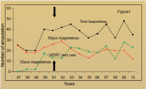
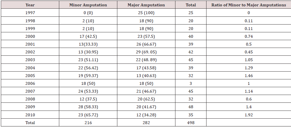
Between 1997 and 2010, we were able to identify a total of 498 lower limb amputations in diabetic patients were performed. Of these 498 amputations, 282 were major amputations Table 1. Results in Table 1 demonstrated a clear reduction in the proportion of major to minor amputation over this study period. The ratio of minor to major amputation rose from 0% in 1997 to 192% in 2010. The number of minor amputations and total amputation rose but the number of major amputations reduced. The effect of introduction of foot clinic in 2001 was clear in this trend Figure 1. When the early group (1997 to 2001) was compared to late group (2006 to 2010) there was significant increase in minor amputations, but no change in major or total amputations. There was significant increase in ratio of minor to major amputations Table 2. The number of newly diagnosed diabetes increased significantly around 2003 in the UK as general practitioners were given incentives to identify undiagnosed diabetes. As a result, amputation rate per 1000 diabetes population would not give correct picture. Therefore, we analysed amputation rate per 100,000 population. The population of area covered by Lancashire Teaching Hospital increased from 333,900 to 356500 between 2001 to 2011. The major annual amputation rate per 100,000 people reduced from 6.6 to 5.1 but minor amputation rate rose from 2.0 to 5.9 between the early and late groups.

Figure 1: Incidences of major, minor and total amputation per 100,000 general populations. The black arrow indicates the introduction of the MDFC at Lancashire Teaching Hospitals NHS Foundation Trust.
Lupinepublishers-openaccess-journal-diabetes-obesity

Table 1: Minor and major amputation rates and Minor: Major amputation ratio 1991-2010. Figures in () represent proportion of amputations that year distributed between major and minor amputations.
lupinepublishers-openaccess-journal-diabetes-obesity

Table 2: Mean annual amputation rate (standard deviation) per year in ‘Early’ (1997–2001) and ‘Late’ (2006–2010) cohorts showing significant rise in minor amputations.

lupinepublishers-openaccess-journal-diabetes-obesity

Discussion

The development of the multi-disciplinary foot clinic has demonstrated that the multidisciplinary approach to diabetic patients can lead to earlier and better recognition of diabetic foot complications. This can lead to prompt and effective referral to the appropriate specialist for swift management. Management of diabetic lower limb complications varies nationally [12] depending on vascular experience, variability in regional protocols and we are hopeful that this approach will encourage a consensus on the management of these complex patients within the Lancashire Vascular Tertiary Referral Centre. The multi-disciplinary approach may have led to change in attitudes around the detection and management of diabetic lower limb complications. The increase in number of diabetic patients following QOF introduction in 2003 without any significant increase in population slightly affected the way we expressed our data. In order to be more representative, we favored the incidence of amputation being expressed as per 100,000 general populations, in order to avoid bias change based on increasing in diabetes prevalence. Despite the observed increasing diabetic population, the proportion of them requiring major amputation is decreasing which is reassuring that our approach is having the desired effect. We noticed increase in minor amputations. This is because same patient could have 5 minor amputations before undergoing major amputation of a leg. Each minor amputation would have prevented one major amputation. However major amputations can be delayed but not totally prevented in many cases as seen by surge in major amputation after few years gap.
Researchers reporting in a study performed in Ipswich Hospital in 2008 found very similar results following the introduction of a multi-disciplinary foot team [2]. This suggests that, irrespective of location, population and other associated risk factors they might harbour, a team-based approach to the care of these high risk patients can result in dramatic improvements in long term limb preservation. The study does have several limitations. The study was performed retrospectively which can mean that the data is less accurate as it relies on coded data which may have been entered incorrectly. The management of diabetic lower limb complications is also affected by the variability in individual surgeon practice. Some surgeons are very aggressive in their limb preservation while others are more pragmatic, and this will affect the outcome.

Conclusion

These findings support the suggestion that a multi-disciplinary approach to these high-risk patients plays an important role in the reduction of lower limb major amputation rates. Over the study period there have been improvements in the vascular, radiological and microbiological management of patients. These disciplines have improved their ability to communicate and work as a multidisciplinary team which will have improved the management of diabetic related complications. Early detection of such complications may allow limb preservation or limb-length preservation over time and we have clearly demonstrated significant improvement in outcomes for one of the most feared and most costly complications for diabetic patients. Improvement in foot care through a multidisciplinary approach and continuous assessment of practice can only ensure long term outcomes will mirror those found in this 13- year study period.

For more Lupine Publishers Open Access Journals Please visit our website:
http://lupinepublishers.us/


For more Open Access Journal on diabetes and Obesity articles Please Click Here:
https://lupinepublishers.com/diabetes-obesity-journal/

Wednesday, October 28, 2020

Tuesday, October 27, 2020

Friday, October 23, 2020

Lupine Publishers| Pharmaceutical Potential of Cerium Oxide Nanoparticles as Anti-Obesity and Anti-Diabetic Nano-Drug

 Lupine Publishers| Pharmaceutical Potential of Cerium Oxide Nanoparticles as Anti-Obesity and Anti-Diabetic Nano-Drug

Lupine Publishers| Diabetes and Obesity

Opinion

Obesity is a universal pathological condition that affected human health seriously. It would be better to say that, treatment methods of obesity have not been effective to date [1]. This problem evoke researchers into a great challenge to overcome it [2]. Broadly speaking, the reactive oxygen species (ROS) play an important role in lipid accumulation [3]. Oxidative stress is an indispensable phenomenon for the adipocyte accumulation [4-6]. Diabetes mellitus (DM) is an endocrine-metabolic disorder that is increasing worldwide due to population aging, urbanization and obesity. Above all, it causes accelerate mortality rate. Increased oxidative stress plays an important role in the development and progression of diabetes and its complications. Diabetes is usually caused by increased production of free radicals or impaired antioxidant defense [7]. Lorcaserin is a serotonin 5-HT2C receptor agonist which imitate from the serotonin effects and causing an increase of satiety and the reduction of the appetite [8]. Qsymia is a combination of the two drugs phentermine and topiramate, which has the role of weight loss by suppressing the appetite and increasing the sense of satiety [9]. It is also worth mentioning that both of these drugs show considerably side effects like dizziness, headache, insomnia, and risk of teratogenicity [10]. Another strategy to decrease body weight is the assumption of dietary polyphenols (such as green tea, resveratrol, curcumin, etc.), that exhibit antioxidant and anti-inflammatory effects related to lipid accumulation [11], but unfortunately, they are rapidly metabolized by enzymes. As a matter of fact they have low stability and bioavailability after the ingestion [12].

Table 1: chemical properties and clinical application of CONPs.

lupinepublishers-openaccess-journal-diabetes-obesity

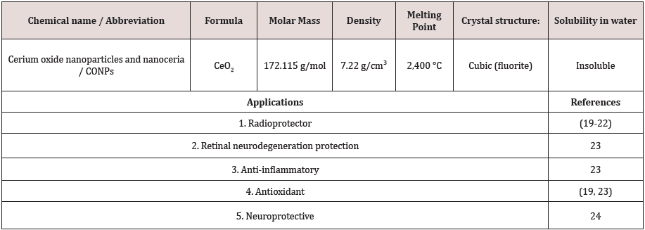
Beneficial effects of cerium oxide nanoparticles (CONPs - CeO2) or nanoceria cover a wide area of applications, ranging from macular degeneration to cancer therapy which listed in Table 1 [13]. CONPs mimic superoxide dismutase and/or catalase activity, depending on the presence of crystalline defects on their surface (Ce3+/Ce4+ ratio) and the environment pH where they accumulate [14]. Nanoceria as Reactive Oxygen Species (ROS) scavenger can self-regenerate their antioxidant properties by switch between the two oxidation states of cerium [15]. Thereby cerium oxide nanoparticles could overcome most of the typical limitations of traditional anti-oxidant agents because of the self-regenerating catalytic properties. Conversely to commercially available drugs against obesity, CONPs have useful advantage to strongly scavenge the ROS production for a long-sustained period of time, thus for one thing it reduces the needed doses and eventually diminish the adverse side effects of other drugs [1]. Strategies to reduce the formation of oxidative stress are important in the treatment of DM [16]. It seems that CeO2 nanoparticles as powerful antioxidant with free radical scavenging properties, is suitable for this purpose [17]. CONPs were thought to increase antioxidant power due to their catalytic effect in stimulating superoxide Dismutase (SOD) activity and detoxifying free radicals by staying active in the tissues for a long time through the spontaneously movement between the oxidized and reduced state [18-24]. It was shown in animal model that CONPs could reduce body weight effectively [1]. These promising results may provide a novel treatment in the clinical setting in the future. Bearing in mind that future studies should scrutinize the biocompatibility and bioactivity mechanism of the CONPs in diabetic patients.

For more Lupine Publishers Open Access Journals Please visit our website:
http://lupinepublishers.us/


For more Open Access Journal on diabetes and Obesity articles Please Click Here:
https://lupinepublishers.com/diabetes-obesity-journal/


Lupine Publishers: Lupine Publishers | Lasers & Pedodontics

Lupine Publishers: Lupine Publishers | Lasers & Pedodontics:  Lupine Publishers | Journal of Pediatric Dentistry Introduction The medical terms such as magical...

Thursday, October 22, 2020

Wednesday, October 21, 2020

Saturday, October 17, 2020

Lupine Publishers| Nutrition and Pressure Ulcers: An Opportunity to Accelerate the Healing Process

 Lupine Publishers| Nutrition and Pressure Ulcers: An Opportunity to Accelerate the Healing Process

Lupine Publishers| Archives of Diabetes and Obesity

Abstract

Pressure ulcers, also known as bedsores, are a prevalent and debilitating condition affecting up to 23% of patients in nursing homes or acute hospital settings [1-3]. Inadequate healing of pressure ulcers increases a patient’s susceptibility to life-threatening infections and prolonged hospital stays, leading to increased healthcare expenditure. Therefore, interventions to prevent ulcers or accelerate ulcer healing are essential to improve patient outcomes and reduce the burden of pressure ulcers on healthcare systems.

Introduction

Pressure ulcers are associated with life-threatening complications and high healthcare costs

Pressure ulcers are often caused by a local breakdown of soft tissue as a result of compression between a bony prominence and external surface causing localized tissue damage [3]. An ordinally, older patients, malnourished patients, patients with low mobility and patients with poor sensory perception are at greater risk of pressure ulcers [3,5]. 70-73% of those who develop pressure ulcers are over 6S years old and 25.16% in Hong Kong nursing homes [3]. Typically, pressure ulcers do not heal well, and inadequate healing increases a patient’s susceptibility to life threatening infections, including sepsis and cellulitis [4-6]. Additionally, poor ulcer healing negatively affects the clinical prognosis of comorbid conditions. Mortality rates in patients with pressure ulcers admitted to nursing homes can be as high as 50% after 1 year compared with 27% in patients without pressure ulcers [1]. Therefore, the cost of treating pressure ulcers is high, ranging from £958-£11,606 per patient in the United Kingdom, depending on ulcer severity [7]. Approximately 90% of this cost is a result of increased nursing care, which includes patient monitoring and wound dressing, but antibiotic Costs for treating infections also contribute [7].

Malnutrition is an independent risk factor for pressure ulcers

Malnutrition has been identified as an independent risk factor for both the development of pressure ulcers and delayed healing [8,9]. Additionally, patients with pressure ulcers have high energy expenditure due to the increased nutritional requirements associated with tissue regeneration and an activated immune response [10]. Figures have also been highlighted by Barker et al. [5], approximately 40% of patients in the acute hospital setting are malnourished [5], pin-pointing the need for nutritional assessment upon hospital admission to reduce the risk of complications. The importance of nutrition has been recognized in the National Pressure Ulcer Advisory Panel (NPUAP)/European Pressure Ulcer Advisory Panel (EPUAP)/ Pan Pacific Pressure Injury Alliance (PPPIA), which recommend assessment of nutrition- al status in patients with, or at risk of developing, pressure ulcers. The guidelines. additionally recommend prescription of a high-energy (30-3Skcal/kg body weight/day), high-protein (1.2S-1.5g/kg body weight/day) diet for patients who are malnourished, or at risk of becoming malnourished, that also includes the importance of amino acids such as arginine and micronutrients [11].

A specific high-energy, high-protein, arginine and micronutrient- enriched oral nutritional supplementation enhances pressure ulcer healing

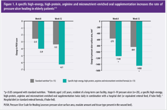
Nutritional support, mostly in the form of high-energy, highprotein oral nutritional supplement can significantly reduce pressure ulcer incidence by 25% in at risk patients compared with standard care [3]. In clinical studies, oral nutrition with specific oral nutritional supplementation enhanced wound healing in patients who were elderly [15] or malnourished [12]. In a 12-week randomized controlled trial, 28 elderly patients with grade II-IV pressure ulcers were randomized to receive a specific high-energy, high-protein, arginine and micronutrient-enriched oral nutritional supplementation twice daily (nutrition formula provided of a total of 500kcaI, 34g protein, 6g arginine, TOOmg vitamin C, and 18mg zinc) in combination with a standard hospital diet (if tube fed, an 1000mL enteral feed with 20% of energy from protein enriched with arginine, zinc and vitamin C [100 kcal, S.S g protein, 0.85 g arginine, 38 mg vitamin C, 2.0mg zinc per 100mL]) or standard nutrition (hospital diet; if tube fed, standard enteral formula comprising 16% of energy from protein without any additional supplement [100kcaI, 4.0g protein, 0g arginine, 10mg vitamin C, 1.2mg zinc per 100mL]). The type and amount of food consumed by each patient was recorded on a clinical register and the mean intake over 3 days standardized to patient weight. Regression analyses were performed to identify the contribution of a specific supplement to any improved outcomes [15]. Results have shown supplementing patients with a high-energy, high-protein, arginine and micronutrient-enriched formula significantly reduced pressure ulcer Scale for healing (PUSH) Score by 85% after 12 weeks (Figure 1; assessment of ulcer surface area, amount of exudate and tissue type), indicating an elevated wound healing with a specific highenergy, high-protein, arginine and micronutrient-enriched oral supplementation. Addition- ally, the specific supplementation was associated with a significant 100% reduction in ulcer surface area after 8 weeks compared with standard nutrition (Figure 1) [15].

Figure 1:

Lupinepublishers-openaccess-journal-diabetes-obesity

Figure 2:

Lupinepublishers-openaccess-journal-diabetes-obesity

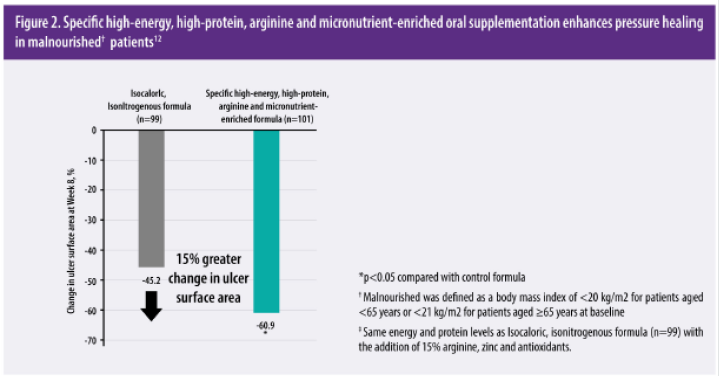
These results have been validated in a large(fi=200), multicenter, randomized controlled trial. The Oligo Element Sore Trial (OEST) reported that the specific oral supplementation significantly enhanced pressure ulcer healing in patients with stage II-lV who were malnourished compared with an isocaloricisonitrogenous formula [12]. Twice-daily supplementation with specific high-energy, high-protein, arginine and micronutrientenriched formula for 8 weeks led to a significantly greater mean reduction in pressure ulcer area (Figure 2) [12].

Management of a sacral pressure ulcer patient by oral supplemented with a specific high protein arginine and micronutrient enriched formula

Introduction This

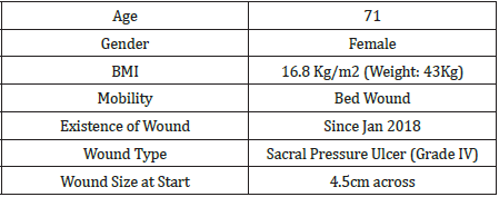
case study describes the management of a patient who developed a sacral pressure sore (Grade IV) during stay in the hospital secondary to hematuria and further care once discharge was followed up in a elderly nursing home setting. Oral Nutritional Supplement was given to promote healing of the wound by Supplementing patient with a high energy, high protein, arginine and micronutrient enriched formula aim promote wound healing.

Treatment regimen

Signs of dead tissue developing within the ulcer with wound on measuring approx 4.5cm across. Ulcer was washed with iodopovidone antiseptic solution and new dressing was applied every day. On 27th Jan 2018 patient was referred to dietitian with past medical history of dementia. Aim was set to promote wound healing and nutritional status as current weight was noted at 43kg and a BMI of 16.8 kg/m2. A high energy, high protein oral nutritional supplementation was recommended twice daily (nutrition formula provided of a total of 500kcal, 34g protein, 6g arginine) in addition to their regular diet and standard wound care and was followed up weekly (Table 1).

Table 1: Patients Characteristics.

lupinepublishers-openaccess-journal-diabetes-obesity

Result

After one week the ulcer had reduced significantly in size (3cm) and new flesh and skin was noticed to be developing. Appetite was slowly picking up and supplementation was adjusted to once daily (nutrition formula provided of a total of 250kcal, 17g protein, 3g arginine). Weight was noted staple whilst ulcer continue to heal. By March 2018, the ulcer was almost healed, and patient’s appetite has returned to normal.

Conclusion

Pressure ulcers are a major burden on health resources, with inadequate healing potentially leading to life threatening infections and an increased risk of morality [1,4,5]. Nutritional supplementation with specific high energy, high protein, arginine and micronutrient- enriched oral formula a significantly reduced pressure ulcer incidence and enhanced wound healing in patients who were elderly [15] or malnourished [12] Figure 3.

Figure 3:

Lupinepublishers-openaccess-journal-diabetes-obesity

For more Lupine Publishers Open Access Journals Please visit our website:
http://lupinepublishers.us/


For more Open Access Journal on diabetes and Obesity articles Please Click Here:
https://lupinepublishers.com/diabetes-obesity-journal/

To Know More About Open Access Publishers Please Click on Lupine Publishers

Follow on Linkedin : https://www.linkedin.com/company/lupinepublishers

Follow on Twitter   :  https://twitter.com/lupine_online

Wednesday, October 14, 2020

Tuesday, October 13, 2020

Monday, October 12, 2020

Saturday, October 10, 2020

Lupine Publishers| Insulin Resistance is Immediately Reduced After Hemodialysis Sessions in Diabetic and Non- Diabetic Patients with End Stage Renal Disease

 Lupine Publishers| Insulin Resistance is Immediately Reduced After Hemodialysis Sessions in Diabetic and Non- Diabetic Patients with End Stage Renal Disease

Lupine Publishers| Diabetes and Obesity

Abstract

Background: Insulin resistance in subjects with End Stage Renal Disease (ESRD) is a strong independent predictor of cardiovascular death. The immediate effect of hemodialysis Session on the state of insulin resistance in patients with ESRD is not very clear and requires further studies.

Methods: This self-control study was carried out at the Alexandria Main University Hospital; the study included 100 subjects with ESRD on regular hemodialysis (50 subjects with type 2 diabetes mellitus and 50 subjects without diabetes mellitus). Fasting plasma glucose, Fasting serum insulin, Homeostasis Model Assessment of Insulin Resistance (HOMA-IR), blood urea, serum creatinine and arterial bicarbonate were assessed before and after the hemodialysis sessions.

Results: The mean HOMA-IR was 4.58 in subjects with diabetes mellitus and 2.37 in subjects without diabetes mellitus. The levels decreased significantly to 1.28 in subject with diabetes mellitus (P< 0.001) and decrease to 0.7 in subject without diabetes mellitus (p< 0.001) after the hemodialysis session. There was a significant negative correlation between HOMA-IR and serum creatinine levels before or after the hemodialysis sessions.

Conclusion: Hemodialysis sessions may cause am immediate improvement in the insulin resistance state in patients with ESRD with or without history of T2DM; this could have some important cardiovascular and metabolic implications on this group of patients

Keywords:Insulin Resistance; End Stage Renal Disease; HOMA-IR

Abbreviations: BMI: Body Mass Index; ESRD: End Stage Renal Disease; HOMA-IR: Homeostasis Model Assessment of Insulin Resistance; T2DM: Type 2 Diabetes Mellitus

Introduction

Diabetic nephropathy is one of the most serious complications of diabetes mellitus; Diabetic kidney disease is responsible for about 40 % of cases with ESRD [1-3]. Insulin resistance refers to decreased insulin ability to exert its physiological actions on different insulin sensitive tissues including the muscles, liver and the adipose tissue. Insulin resistance is not only related to the pathogenesis of type 2 diabetes mellitus (T2DM) but may also have a role in the development of chronic kidney disease [4]. In general, insulin resistance could be a pre receptor resistance, a receptor resistance or a post receptor resistance. Insulin resistance in chronic renal disease is mainly related to a post binding abnormality in insulin action [5-8]. Insulin resistance may lead to kidney damage through altering the renal hemodynamics by mechanisms such as activation of the sympathetic nervous system, sodium retention and decreased Na+, K+-ATPase activity [9]. HOMA-IR can be used as an alternative to the Hyperinsulinemic Euglycemic Glucose Clamp to estimate insulin resistance in patients with chronic kidney disease [10-13]. The effect of hemodialysis sessions on the state of insulin resistance in patients with ESRD was not clear. The aim of this study was to assess the immediate effect of hemodialysis session on insulin resistance in patients with ESRD.

Materials and Methods

This self-control study included 100 adult patients with ESRD on regular hemodialysis (more than 6 months, three times per week, 4h session duration) selected from the Alexandria main university hospital. On the other hand, patients with morbid obesity (BMI >40 kg/m2), Under-weight (BMI<18.5kg/m2), type 1 diabetes mellitus or decompensated liver cirrhosis were excluded. The studied group was equally divided in to two subgroups; subgroup I (50 subjects with T2DM) and subgroup II (50 subjects without T2DM). The study was conducted in accordance with the Declaration of Helsinki and was approved by the Ethics Committee of Alexandria University, Egypt. Signed informed consent was obtained from each subject before any study-related activities [10]. Patients were asked to fast for 8 hours before HD session and to continue fasting till the end of the session. Blood urea, serum creatinine, arterial bicarbonate, fasting plasma glucose and fasting serum insulin were measured just before hemodialysis session and repeated immediately after hemodialysis session. Insulin resistance was calculated by HOMA IR using the formula: Fasting serum insulin (μUml) x fasting plasma glucose (mmol/L)/ 22.5 [10]. Data was analyzed by using student ‘t’ test (paired and unpaired) and Pearson’s correlation coefficient (r).

Result

Descriptive Statistical Data of the Studied Patients

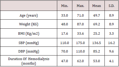
The study included 100 patients with ESRD, the mean age of the studied patients was 49.7 years, the mean BMI was 25.2 and the mean duration for which the patients were on regular hemodialysis was 53 months (Table 1).

Table 1: Descriptive statistical data of the studied patients (n=100 patients).

lupinepublishers-openaccess-journal-diabetes-obesity

Blood Urea

The mean of blood urea levels significantly dropped from 101.2 mg/dl before hemodialysis sessions to 62.7 mg/dl after the hemodialysis sessions (p=0.013).

Serum Creatinine

The mean of serum creatinine levels significantly dropped from 10.0 mg/dl before hemodialysis sessions to 6.1 mg/dl after the hemodialysis sessions (p=0.011).

Arterial Bicarbonate

The mean of arterial bicarbonate levels significantly increased from 19.5 mmol/l before hemodialysis sessions to 24.5 mmol/l after the hemodialysis sessions (p=0.038).

Fasting Plasma Glucose Level

a) In subgroup I, the mean of fasting plasma glucose levels was 127.38 mg/dl before hemodialysis sessions and 96.62 mg/dl after the hemodialysis sessions (p=0.001<).

b) In subgroup II, the mean of fasting plasma glucose levels was 88.48 mg/dl before hemodialysis sessions and 78.96 mg/dl after the hemodialysis sessions (p=0.001<).

Fasting Serum Insulin

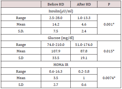
Table 2: Fasting serum insulin level, fasting glucose level and HOMA IR before and after hemodialysis in the studied patients (n=100 patients).

lupinepublishers-openaccess-journal-diabetes-obesity

a) In the 100 studied patients, the mean of fasting serum insulin levels significantly dropped (p=0.001) from 14.2 μU/ml before hemodialysis sessions to 4.6 μU/ml just after the hemodialysis sessions (Table 2).
b) In the subgroup I, the mean of fasting serum insulin levels significantly dropped (p=0.001) from 17.3 μU/ml before hemodialysis sessions to 5.45 μU/ml just after the hemodialysis sessions.
c) In the subgroup II, the mean of fasting serum insulin levels significantly dropped (p=0.001) from 11.18 μU/ml before hemodialysis sessions to 3.64 μU/ml just after the hemodialysis sessions.

HOMA IR

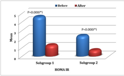
In the 100 studied patients, the mean of HOMA IR levels significantly dropped (p=0.0074) from 3.5 before hemodialysis sessions to 1.0 just after the hemodialysis sessions (Table 2). In comparison between the two studied subgroups, in subgroup I, HOMA IR before (HD) ranged between 0.94-16.32 with mean value 4.58±3.37 and in subgroup II ranged 0.58-5 with mean value 2.37±1.16. While in subgroup I, HOMA IR after (HD) ranged 0.71- 3.76 with mean value 1.28±0.66 and in subgroup II ranged 0.17- 1.70 with mean value 0.70±0.37. There was statistical significant decrease in HOMA IR just after the hemodialysis sessions in both subgroups (P < 0.05) (Figure 1). There was a negative significant correlation between HOMA IR before hemodialysis sessions and creatinine level before hemodialysis sessions (r = -.225, p = .024), also there was a negative correlation between HOMA IR levels after the hemodialysis sessions and creatinine levels just after the hemodialysis sessions (r = -.286, p = .004).

Figure 1: Comparison between the two studied subgroups regarding HOMA IR.

Lupinepublishers-openaccess-journal-diabetes-obesity

Discussion

Insulin resistance is a characteristic feature of ESRD. Different mechanisms are implicated in the pathogenesis of insulin resistance in ESRD including; low physical activity levels, toxins of the uremic state, metabolic acidosis, electrolyte disturbance, chronic inflammatory state, and abnormal adipokines levels leading to a post insulin-receptor defect in different insulin sensitive tissues [14,15]. In this present study, there was a significant decrease in fasting serum insulin levels after hemodialysis in the studied patients regardless of whether they suffer from T2DM or not; this was previously reported by Nad, et al (2014) who found that the serum insulin levels significantly decreased after hemodialysis in the diabetic and non-diabetic patients [16]. The main fining of our study was the significant improvement in IR immediately after the hemodialysis sessions even in patients without T2DM. Duncan E et al (2009) found HOMA-IR levels in the diabetic group significantly decreased after HD, but there was no significant change in levels of HOMA-IR in their non-diabetic participants [17]. Another important fining of our work is the significant negative correlation between HOMA-IR and serum creatinine levels. Similarly, Kobayashi S et al (2005) found that insulin resistance is correlated linearly with decline in renal functions [1].

Conclusion

Hemodialysis sessions may cause am immediate improvement in the insulin resistance state in patients with ESRD with or without history of T2DM; this could have some important metabolic implications on this group of patients.

For more Lupine Publishers Open Access Journals Please visit our website:
http://lupinepublishers.us/


For more Open Access Journal on diabetes and Obesity articles Please Click Here:
https://lupinepublishers.com/diabetes-obesity-journal/
To Know More About Open Access Publishers Please Click on Lupine Publishers

Follow on Linkedin : https://www.linkedin.com/company/lupinepublishers

Follow on Twitter   :  https://twitter.com/lupine_online


Friday, October 9, 2020

Thursday, October 8, 2020

Wednesday, October 7, 2020

Tuesday, October 6, 2020

Monday, October 5, 2020

Friday, October 2, 2020

Lupine Publishers| Semaglutide versus liraglutide for treatment of obesity

  Lupine Publishers| Journal of Diabetes and Obesity Abstract Background: Once weekly (OW) semaglutide is a glucagon-like peptide...